Imaging Laboratory
Imaging Laboratory of the Jagiellonian |Centre of Innovation offers a broad range of services within the scope of surface analysis, based mostly on Raman spectroscopy and electron microscopy.
Raman spectroscopy (Raman effect)
Raman spectroscopy is one of the most versatile analytic techniques used to determine the chemical composition of the studied material. The basis for this technique is non-elastic scattering of monochromatic light. The energy of non-elastically scattered light is different than the energy of the incident light. The recorded change in energy may be presented as a Raman spectrum characteristic for the given substance.
The Laboratory is equipped with two state-of-art Raman spectroscopes:
WITec alpha 300 RSA+ and alpha 300 R, equipped with laser lines with wavelengths of 532, 633 and 785 nm. The devices are built based on a confocal microscope which allows accurate measurements in the chosen plane of the sample, as well as 2D and 3D Raman mapping. Moreover, the spectrometers have a profile meter (TrueSurface®) which allows mapping of the sample surface upon measurement. Conventional air lenses are supplemented with an immersive lens; hence measurements can be performed also in aqueous solutions. Additionally, WITec alpha 300R device was equipped with 90- and 45-degree polarizers, as well as circular polarization for the 532 nm line, as well as specialist software Particle Scout for particle size analysis.
Measurements using both spectrometers do not require previous tagging and are non-destructive to the studied materials
We offer:
- Determination of chemical composition of the studied material based on comparison with reference substance spectra
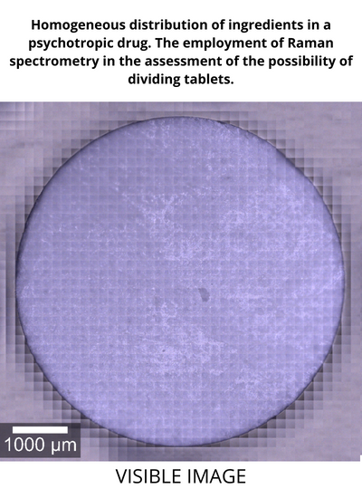
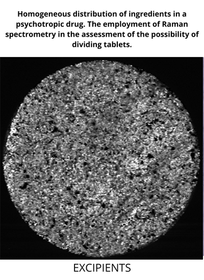
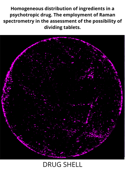
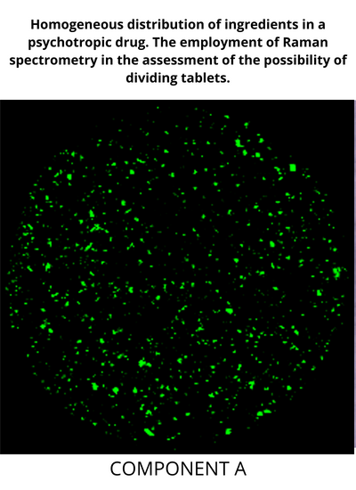
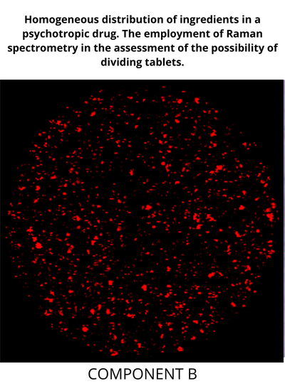
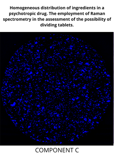
- Measurements within the scope of imaging of distribution of chosen substances in tablets (spatial resolution up to 200–300 nm)
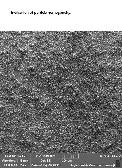
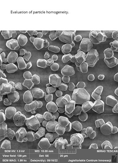
- Homogeneity testing for solid formulations
- Sample topography and confocal measurement analysis
- Measurements of chemical compounds to allow discrimination of their polymorphic forms
- Testing permeability of semi-liquid formulations through the skin as a function of exposure time and conditions
- Measurements of polymeric materials
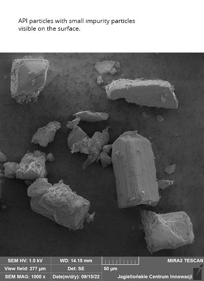
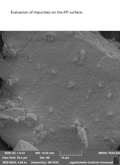
- Possibility to identify impurities in solids
- NEW – particle size measurement – simultaneous identification and classification of the substance in the sample, and analysis of particle size distribution for powdered samples and suspensions
- NEW – measurements using immersive lens – possibility to measure biological samples (cells, tissues) and other samples suspended in aqueous solutions
- NEW – possibility to perform surface-enhanced Raman spectroscopy (SERS) measurements
- NEW – application of polarization measurements for measuring polymorphic forms and determination of biomolecule orientation in biological samples
Additionally, the Raman spectrometer may be used for:
- Study the chosen tissue fragments
- Perform cell testing in vitro with ability to differentiate cell organelles
- Preform chemical imaging without histological staining
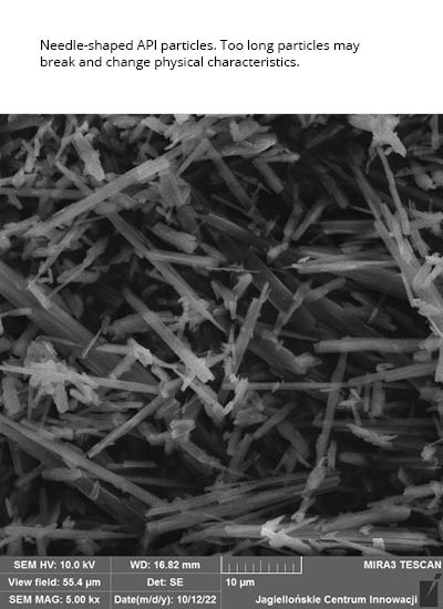
- Test medicinal substances for active substance distribution, determination of crystallization degree and presence of defects such as inclusions or breaks
- Characterize the components of painting layers (pigments, fillers, adhesives)
- It is possible to study semiconductor surfaces
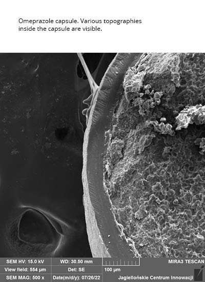
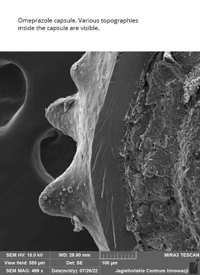
Scanning electron microscopy
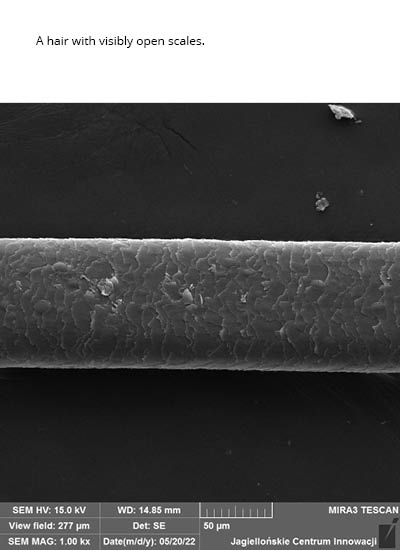
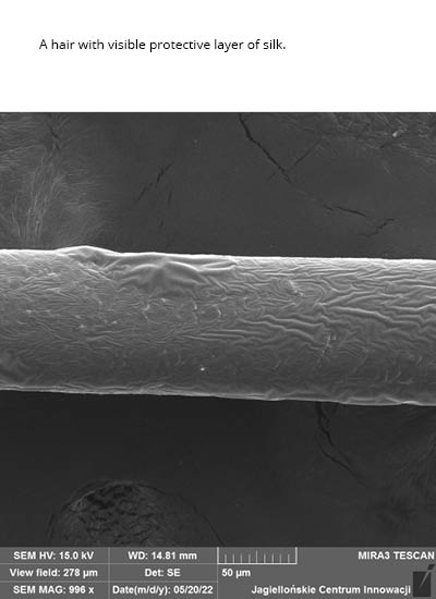
Scanning electron microscopy allows studying conductive materials (metal oxides, metals and their alloys), as well as non-conductive materials such as cells, tissues, medications (active substances and excipients), polymers and also ceramic, composite and organic materials.
The Laboratory owns a scanning electron microscope (Mira3-FEG-SEM, Tescan) with field emission (Schottky emitter), equipped with SE, BSE, and LVSTD detectors, and X-ray energy dispersion spectrometer EDX (X-Act Oxford Instruments), as well as a cooling table (Peltier’s cell) operating in the temperature range from -30°C. The microscope allows working in high, low and variable vacuum.
We offer:
- Imaging of conductive and non-conductive samples in high and variable vacuum conditions
- Analysis of morphology and chemical composition of non-conductive samples in low and variable vacuum
- Qualitative and semi-quantitative analyses of elements with atomic number Z≥5 – surface, point, line measurements as well as element distribution map in micro-areas using the EDX spectrometer EDX
- Analyses of biological samples in situ, observation of dynamic changes such as hydration or dehydration of sample, and possibility to observe the phase transitions (Peltier’s table)
- Observations of interactions between the sample and its changing environment
Active, remote participation in SEM measurements is possible via MS Teams, YouTube, Google Meet or other platforms.
Head of the Imaging Laboratory
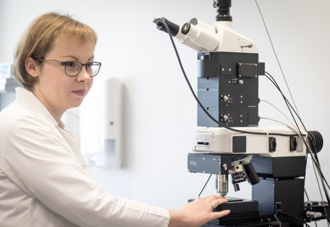
Emilia Staniszewska-Ślęzak, PhD
Emilia Staniszewska-Ślęzak, PhD, graduated from the Faculty of Chemistry of the Jagiellonian University. For over 7 years, she has been interested in Raman and absorption spectroscopy in the infrared range. A co-author of many scientific publications showing the application of the above-mentioned techniques in biomedical and art conservation research. She has been connected with the Jagiellonian Center of Innovation since 2017 and she is responsible for performing research with use of Raman spectroscopy.
For a quote, please contact:
Diana Dołęga, PhD
Mobile: +48 517 062 813
E-mail: diana.dolega@jci.pl
Our clients


Application notes
dr Emilia Staniszewska-Ślęzak
Distribution of the active substances in the tablet assessed with topographic raman imaging
More Show all